Tag Archives: dissection

Rat Dissection


rat_dissection

Today we planned to do a rat dissection, but due to the frosty state of our subjects, we have postponed this investigation until Tuesday. In the meantime, we will answer the questions we can, using digital resources:

Heart and Kidney Dissections

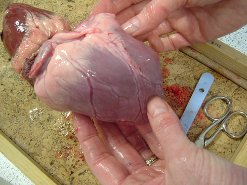
cow's heart

This image shows a cow’s heart ready for dissection – note the diagonal line which separates the left and right sides of the heart – thin-walled atria at the top and thicker walled ventricles at the bottom. On Friday this week we will also be dissecting sheep’s kidneys, to show renal arteries, medulla and cortex.  This great interactive animation shows how water, glucose and salts area reabsorbed and how urea is excreted by the kidneys. This site has five different sections showing how the nephron, capsule, proximal, loop and duct work – make sure you find them all. How our kidneys work from Kidney Health Australia. Kidneys Explained  from the Better Health Channel.

You are in for a treat on Monday – I have been able to get a pig’s heart, liver, lungs and kidneys from a local farmer. Pig tissues are sometimes used in humans, as human transplants are not always available and pig organs are about the same size and able to be produced in large numbers. Studies at Cambridge University found that people who receive transplanted pig organs are unlikely to contract incurable, infectious diseases. Scientists at Melbourne’s Alfred Hospital have kept pig’s lungs alive and functioning with human blood. The pigs used have been genetically modified to reduce blood clotting and the chances of rejection in humans. This practise raises serious ethical issues that must be carefully considered by medical practitioners, governments and society. There is the possibility of bringing animal diseases into the human population, peoples attitudes to ‘part-human part-pig’ organisms, as well as the argument that humans should not be tampering with ‘god’s  work’. Leave me a comment and let me know your thoughts about xenotransplantation – animal-to-human transplants.

wild boar kidney

Transport in Animals

Image Source

This week we start to look at the circulatory, respiratory and excretory systems of animals and the xylem and phloem cells in plants. We know now how different organisms obtain their nutrients, now we need to know how nutrients and oxygen get to every cell in the body and how wastes are removed from it.

Great animation about how the heart works here. Video of heart and circulatory system here.

Human Anatomy Online – interactive diagrams of all the systems, including nervous, skeletal and reproductive. KLB Science Interactivities have produced a clever quiz on the heart and circulatory system. More great Human Body stuff from National Geographic here. Virtual microscope images of the circulatory system from the Indiana University Bloomington.

Image Source – Note the five different types of white blood cells.

Components of blood

Virtual microscope slide of blood

Virtual microscope images of arteries, veins and capillaries showing tissue types.

RESPIRATORY SYSTEM

Don’t get confused between cellular respiration and breathing! Cellular respiration is the process that converts glucose and oxygen to energy within the cells. Oxygen is supplied to those cells by the red blood cells, which carry oxyhaemoglobin to cells and remove carbon dioxide from cells. The respiratory system includes the lungs, trachea, bronchioles and alveoli, which carry air into and out of the body.

Respiration – University of Melbourne animation of lung structure showing alveoli.

Habits of the Heart – Lung Structure from the Science Museum of Minnesota.

Video of the Respiratory System

Human Anatomy Animations of the respiratory and circulatory sytems from Bioanime. This site includes animations of all the human tissue types, including the different types of white blood cells.

Rat Dissection

Source: Biology Online

This Wednesday afternoon we will be dissecting laboratory rats, which are specially bred for scientific purposes. This practical exercise is optional, but an excellent way to gain scientific skills of careful observation, identification of body parts and an understanding of the structure and function of the digestive system of mammals. If you plan to continue your science education at university, you will find this a valuable introduction to laboratory dissections. Make sure you read the practical instructions thoroughly, work slowly and carefully and document your progress with video or a digital camera. Remember that ‘dissection’ does not mean ‘to cut up’, it means ‘to expose to view’ – once something has been cut, it can’t be undone, so know what organ or tissue you are cutting and why.

Rat Dissection – Part 1: Exposing the Abdomen

Rat Dissection – Part 2: The Digestive System (Warning – graphic images)

Rat Dissection – Part 3: Identifying the organs of digestion.

What did you learn about dissection and the digestive system of a mammal? Compared to the length of the rat, how long was it’s alimentary canal? What was the difference in the wall of the stomach and the small intestine? What did you notice about the contents of the alimentary canal as they moved towards the rectum? What surprised you most about the inside of a rat?

Procedure for rat dissection.

Classification of the rat and glossary of terms (dorsal, ventral, thoracic etc.)

Check out Miss Baker’s Biology Blog, “Extreme Biology” for a video of a dogfish shark dissection.