Click on this link to join the Hawkesdale Biology Quizlet class:
Our Quizlet Class
Leave a reply
Click on this link to join the Hawkesdale Biology Quizlet class:
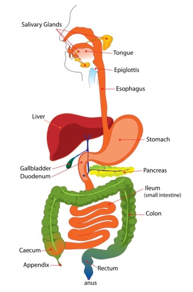
Image Source – Attribution: By Leysi24 (Own work) [CC BY-SA 3.0 via Wikimedia Commons
Firstly, dismantle the human torso model in the science laboratory and describe what you know about each part of the alimentary canal and associated glands and organs. Draw and label a detailed diagram, showing each of the organs above. Then complete the “Cut-and-Paste your Guts” activity, identifying each organ from it’s description and pasting each description into your book, in the order that food would pass through, on it’s 12 hour journey through the 7 metres of the digestive system.
Next, match the skulls (noting the teeth structure and position of eye sockets) with the corresponding herbivore, omnivore and carnivore digestive systems. Describe the diet of each organism, explaining your reasoning in terms of teeth structure, size of stomach and length of intestines, any enlarged organs and corresponding diet.

Unit 1 – Year 11 Biology
Unit 3 – Year 12 Biology

GTAC – Introduction to Photosynthesis on YouTube
GTAC – Photosynthesis – Light Dependent Cycle and an Animation showing six cycles of the Light Independent Cycle on YouTube
Tony, from GTAC, demonstrated a photosynthesis experiment in which equal quantities of spinach leaves were placed in four clear, closed containers. Each container was subjected to light of the same intensity, but one had no filter (control) and the other three were wrapped in coloured cellophane (red, blue and green, as shown above). The coloured cellophane filters out different wavelengths of light, so the red cellophane reflects red wavelengths and allows other wavelengths to pass through. Each container had two probes, measuring oxygen and carbon dioxide concentrations in parts per million (ppm). What would you expect to happen in the cellophane-covered containers compared to the control?
Tony was also able to answer two questions that students have about DNA transcription.
(1) Where does the mRNA molecule go after transcription? “A single mRNA can be translated many times by ribosomes into polypeptides (it’s one way a cellular response dependent on gene expression can be amplified). After that mRNA is degraded, releasing individual nucleotides which can then be recycled into new mRNA. In eukaryotic cells, the mRNA is protected by the 5’ methylguanosine cap and the 3’ poly-A tail. When these are removed from the ends, presumably in response to an intracellular signal that says the mRNA is no longer required, the mRNA becomes susceptible to degradation.”
(2) When and where does transcription occur? “I would say transcription (the process by which the mRNA is first made from DNA template) occurs in the nucleus of eukaryotic cells almost continuously but the genes being expressed change throughout the cell cycle and in response to stimuli. For example, genes relevant to growth may be transcribed during G phases. A special set of genes relevant to DNA synthesis are transcribed during S phase. If a (stem) cell received a differentiation signal, a relevant set of genes would be switched on for differentiation into a particular cell type. I would say the only time transcription ceases is when the chromosomes condense for mitosis and cytokinesis. Essential proteins are still around to ensure cell division proceeds as intended. After cell division and the chromosomes de-condense, it’s back to business as usual.”
Thanks Tony for these valuable extensions to our Year 12 Biology program at Hawkesdale P12 College.
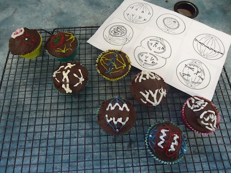
Students in Year 11 Biology are learning the phases of mitosis, so we baked and decorated these cupcakes. Students now have a good understanding of what happens inside the nucleus during:
Watch the Cells Alive Interactive and describe where in each cycle are the three checkpoints that allow DNA replication and mitosis to continue. Why is it incorrect to suggest that the cell is “resting” during interphase, between mitotic cycles?

Both these roses come from the same bush in my garden at home. The one on the left is how the rose normally looks, year after year. This year, on a single branch sprouting to the side, there are about six flowers that look striped, like the one on the right. This article, from the American Rose Society, describes how a genetic mutation can cause this change in pigments.
Stripes may also result from spontaneous or induced mutations. Mutations are sudden changes that occur at a very low frequency in a gene. Spontaneous mutations (popularly known as ‘sports’) alter the existing genes and their expression, resulting in stripes. Induced mutations by irradiation or chemical mutagens also lead to genetically-altered pigmentation, and the result is stripes. Stripes may develop as a result of the transmission of genes responsible for stripes through hybridization. Viral infection that causes variegation in tulips may also cause stripes in roses. These infections could interfere with physiological functions of pigmentation, giving them a striped appearance.
It is possible that a mutation has occurred during mitosis somewhere at the base of the new branch and all the cells in the new branch carry the mutated gene, which is expressed as a striped phenotype. If this is the case, a cutting from this branch will also produce striped flowers. So, I will take a cutting and propagate this rose, to see if we can produce more of these beautiful mutants!
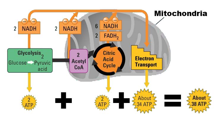
Today our Year 12 students had the opportunity to work with Fran and Catherine from the Gene Technology Access Centre to learn more about cellular respiration. They participated in a 90 minute Polycom session using models and interacting with experts to gain insight into how cells respire and how research into this biochemical pathway may lead to the development of treatments for cancer. It is a wonderful opportunity for small, rural and isolated schools to be able to connect with this world-class educational centre, without the expense and inconvenience of long travel times, permission slips, risk-assessment forms and arranging buses or train tickets. On Monday we will participate in another GTAC session: “The Many Colours of Photosynthesis”. Students’ comments included:
“The experiment that was demonstrated was something we couldn’t do at school, so it was good to learn how that was done and analyse the data.” – Kiri
“The slideshow and worksheets were clear and informative, allowing us to understand and visualize the process of respiration”. – Che
“It was all really good, especially knowing what is important for the exam”. – Stephanie
Quizlet is a website where you can create sets of terms and definitions to assist your learning.
Chapter 1 – Cells – Discovery and Exploration (Britt)
Chapter 2 – Structure and Function of Cells (Georgina)
Chapter 3 – Composition of Cells (Tayla)
Chapter 4 – Cell Replication (Ruby)

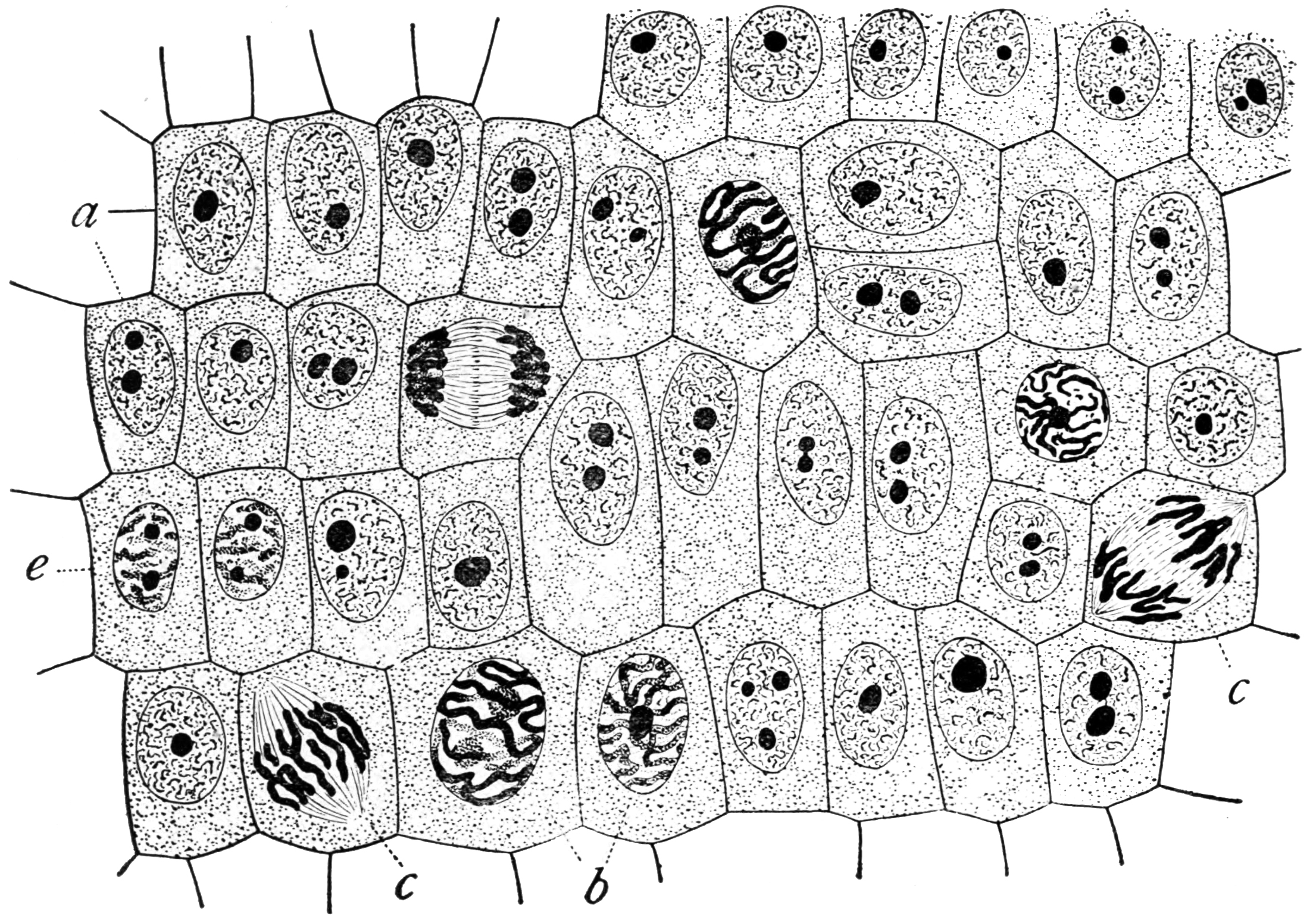
When cells divide during mitosis, each daughter cell contains an identical set of chromosomes to the parent cell. How can this occur? Watch these animations to find out:
The Cell Cycle includes these five phases of Mitosis and Cytokinesis: