Monthly Archives: February 2014

The effects of temperature and pH on enzyme function.


At the Gene Technology Access Centre on Monday we spent the day learning about the structure and function of enzymes. As well as a lecture and practical experiment, we had the opportunity to use a computer program for protein modelling. GTAC has some great online resources for teaching and learning, including this slideshow about Enzyme Action.

Enzymes have specific characteristics:
– enzymes are proteins, made up of amino acids
– enzymes have specific primary, secondary and tertiary structures
– enzymes are specific to substrates
– enzymes are biological catalysts (they speed up a reaction)
– enzymes have optimum temperature and pH ranges
– enzymes are not changed or used up in a reaction
– enzymes have an active site, which is where the substrate is broken down or the products are made
– enzymes can contain co-factors (ions, such as chlorine or calcium) that assist to attract the reactants to the active site

We worked with amylase, an enzyme that breaks starch down into disaccharides. We used iodine to indicate the presence of starch and a photo spectrometer to measure the degree of staining of the medium. The higher the photo spectrometer reading, the more starch, which meant the less enzyme action. We stopped the reaction using an acid, which denatures the enzyme and prevents the break down of starch. Our results showed that the optimum temperature of amylase activity was about 40 degrees and the optimum pH was 6. This is what you might expect from human amylase, which would be working at normal body temperature (37 degrees) and neutral (or slightly acidic) pH in the mouth.

Effect of temperature on cell membranes using beetroot

beetroot-prac-large


Cell_membrane

Image source

In the egg osmosis experiment we used a chicken egg as a model of an animal cell to demonstrate the movement of water across a semi-permeable membrane. We have learned about the different components of the plasma membrane, including the phospholipid bilayer, integral and peripheral proteins, glycoproteins and glycolipids. We know that it is sometimes referred to as a “fluid mosaic” referring to it’s flexible structure of different parts.

Today we are going to test the effect of temperature and solvents on the cell membrane, using cores of raw beetroot. Beetroot is brightly coloured due to the presence of betalain and this coloured pigment can assist us to determine the effect of different temperatures and concentrations of solvents on the cell membrane. When the cell membrane is damaged, the pigment leaks out, so the more damage that occurs, the darker the colour of the medium into which the pigment leaks.

Your task is to design and complete an experiment with an aim, hypothesis, list of materials and equipment, method, results, discussion and conclusion. Your variable can be temperature OR concentration of a solvent (ethanol, for example) or detergent.

Resources:

An egg as a model cell

egg_experiment

Image source

A chicken egg with the shell removed is often used as a model to show how osmosis works – the experiment we did at Federation University, Ballarat, showed how the eggs gain or lose mass depending on the concentration of the solution that they are placed in. It is important to know that the membrane of the egg is not a true biological membrane or plasma membrane. In fact, a chicken egg is a very specialized cell and the membrane is actually composed of keratin fibres – the same protein that makes up human hair, finger nails and rhino horns. Thanks to Andrew Douch for finding this article about chicken egg membranes, with scanning electron micrograph images.

Notice in the image above, the egg in 5% saline solution sinks (indicating that the egg contents are more dense than the solution) and the egg in the 10% saline solution floats (indicating that the egg contents are less dense than the solution). This image should give you a clue as to which egg gains water and which egg loses water by osmosis.

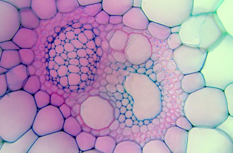
Cell membranes and organelles

plant_cells

Image source

Key knowledge:

  • Understand the extent of the plasma membrane in forming a series of membraneous channels for the packaging and transport of biomolecules throughout eukaryotic cells.
  • Enhance knowledge and understanding of the structure and function of cell organelles.
  • Distinguish the different ways in which biomolecules enter or leave cells.
  • Develop knowledge and understanding of connections between cells.
  • Extend understanding of apoptosis.

Digital resources:

Possible Tasks:

  1. Create a “Thinglink” interactive image, labelling the different parts of a eukaryotic cell.  
  2. Create a database of terms and definitions on Quizlet. 
  3. Decorate a cake to show the different organelles of a plant or animal cell. Label with toothpick flags.
  4. Create a video describing the different forms of active transport using “common craft” style or 3D animation.
  5. Create a labelled model of the “phospholipid bilayer with integral and peripheral proteins”.

Creating model macromolecules

Biology_sweets

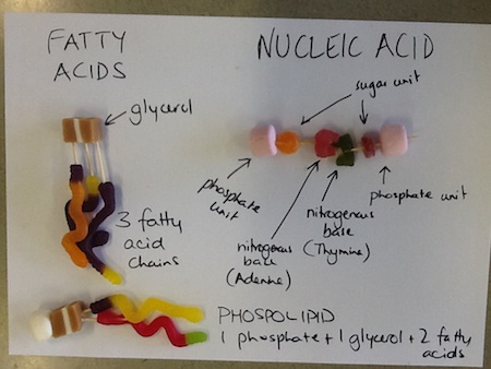
Students enjoyed this class, consolidating their knowledge of carbohydrates, lipids and nucleic acids using soft lollies and toothpicks today. They were able to show that disaccharides are made up of two monosaccharides and polysaccharides are made up of many sugar units (using fruit pastilles) and that lipids consist of a glycerol unit (jersey caramel) and 3 fatty acid chains (jelly snakes). Phospholipids consist of a phosphate group (marshmallow), a glycerol (jersey caramel) and two fatty acid chains. DNA consists of a sugar (fruit pastille) and phosphate (marshmallow) backbone and pairs of nitrogenous bases (jelly joiners), arranged in a double helix. Some showed the two hydrogen bonds between Adenine and Thymine and the three hydrogen bonds between Cytosine and Guanine.

As our next lesson will not be until after the VCE Study Camp, please continue to read through Chapter 2 (Membranes and Cell Organelles), as the practical experiment we are doing at Federation University is all about cell membranes. You will need to understand the following terms and definitions:

  • prokaryotic and eukaryotic
  • apoptosis
  • plasma membrane
  • phospholipid bilayer
  • partially permeable
  • diffusion and osmosis (passive transport)
  • hydrophilic and hydrophobic (or lipophilic)
  • channel-mediated and carrier-mediated
  • active transport
  • phagocytosis, pinocytosis, exocytosis and endocytosis
  • ribosomes, endoplasmic reticulum and golgi complex
  • lysosomes, peroxisomes and endosomes
  • chloroplasts – including lamella, grana and thylakoids

I suggest you create a set of Quizlet flashcards using these terms and definitions.


Biological macromolecules: Nucleic acids

Biomacromolecules – Nucleic acids and proteins (Part A) from GTAC

Biomolecules – Nucleic acids and proteins (Part B) from GTAC

Nucleic acids (DNA and RNA) are the next group of macromolecules that we are looking at. Nucleic acids are made up of monomer units that consist of a phosphate group, a sugar unit and a nitrogenous base. The nitrogenous bases in DNA are Thymine (T), Adenine (A), Guanine(G) and Cytosine (C). In RNA, the Thymine (T) is replaced by Uracil (U). Thymine (or Uracil in RNA) always pairs with Adenine (2 hydrogen bonds) and Guanine always pairs with Cytosine (3 hydrogen bonds).

DNA_Nucleotides

Biological Macromolecules – Lipids

Lipids are fats (solid at room temperature – from animals) and oils (liquid at room temperature – from plants). Next time you are in the supermarket, notice how many different types of fats and oils you can buy, from butter and lard to olive oil and sunflower oil. Triglycerides, cholesterol and phospholipids are all examples of this class of macromolecules, which are characterised by long carbon chains and being organic molecules, insoluble in water.

Biological Macromolecules – Carbohydrates

Last week we started looking at the large molecules that make up all living organisms – carbohydrates, lipids, proteins and nucleic acids. Due to their size, number of monomers and different combinations of monomers and how they are joined, there are huge number of these macromolecules, so they are grouped according to common characteristics. All carbohydrates are made up of Carbon, Hydrogen and Oxygen atoms and have the general formula (CH2O)n. How many carbon atoms do you think triose, pentose and hexose have?

Monosaccharides: glucose, fructose, galactose

Disaccharides: sucrose, lactose

Polysaccharides: chitin (insect exoskeletons and fungi cell walls), cellulose and glycogen