Tag Archives: cells

Mitosis – Cell replication

mitosis in an African globe lily endosperm

Photo Source

This week we have started Chapter 4: Cell Replication, looking at how cells divide for growth, maintenance and repair. Watch the Cells Alive animation that shows the four stages of Mitosis – Prophase, Metaphase, Anaphase and Telophase. The in-between phase is Interphase, when the chromosomes are not visible. What stage is shown in the electron micrograph above? How can you tell?

This site, at NOVA Online, shows how mitosis and meiosis compare. The McGraw-Hill site also has a good animation showing mitosis and cytokinesis (division of cytoplasm and formation of two separate cells).

The most recent edition of New Scientist has an interesting article about how bone cells form –  bone marrow cells can be induced to form bone, fat or blood depending on chemical and physical cues. In an experiment performed at the University of Chicago, scientists induced bone marrow cells to form bone cells in angular moulds (star-shaped or rectangular) and fat cells in curvy moulds (circles and flower shapes).

Heart pumps out new cells!

Image Source

Swedish scientists have used radioactive carbon-14 to show that between 0.5 and 1.0% of heart cells regenerate each year, depending on age. Read more here.

We were talking about bees in class during the last week of term – I happened upon this interesting article from the Scientific American – “Plan Bee: As Honeybees Die Out, Will Other Species Take Their Place?”

I’ve also added a few more resources to the Free Stuff! column on the RHS – Check out “Biology Q and A” for over 1800 questions and answers about Biology.

Quia Quizzes!

Photo Source

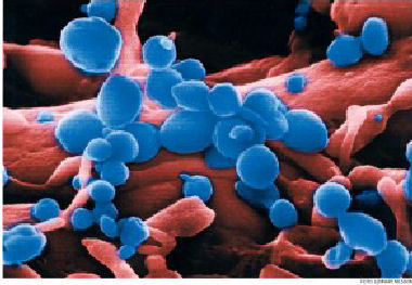
“The first high-resolution close-up photographs of the H5N1 avian flu virus, taken by science photographer Lennart Nilsson, appeared in the Swedish daily Dagens Nyeter (DN) on November 7, 2005, in what the newspaper said was a world exclusive.”

Try these fun activities from Quia to revise Unit 1: Area of Study 1: Cell Structure and Function.

Cell organelles: http://www.quia.com/jg/1624832.html (Word search, Concentration or Flash Cards)

Cell organelles and Photosynthesis review: http://www.quia.com/ba/265821.html (Battleships)

Cells – Who wants to be a millionaire? : http://www.quia.com/rr/450977.html

The cell and plasma membrane: http://www.quia.com/cb/447399.html (Challenge board)

Stages of Mitosis: http://www.quia.com/rd/161026.html (Ordered List)

Revision of Unit 1: Area of study 1: Cells in Action

 
Use My Studiyo to create your own quiz to revise one of the following concepts:

  • Types of cells (prokaryotic, eukaryotic, plant, animal, bacterial)
  • Mitosis
  • Structure and function of cells
  • Biomacromolecules
  • Photosynthesis and respiration
  • Transport across membranes

Make sure you have at least ten questions.

Mitosis in a Lymphoma Cell

Photo Source

This week we are starting Chapter 4: Cell Division. On Friday we watched a Clickview video showing the process of cytokinesis and mitosis, which included a discussion with a cancer surgeon about the types of treatments (chemotherapy and radiation) used to prevent cells dividing and multiplying, as they do in tumours. All living organisms undergo cell division – in unicellular organisms (such as bacteria and yeast) it is how they reproduce. In multicellular organisms, cells divide and multiply for growth, repair and maintenance.

Certain tissues, such as root tips and bone marrow, actively grow and divide and it is relatively easy to find cells undergoing mitosis. We will look at some prepared slides to see the different stages of mitosis – prophase, metaphase, anaphase and telophase – and cells in which the chromatin is not visible (interphase).

Download and print a Mitosis worksheet here.

Another Mitosis worksheet here.

Introduction to Mitosis worksheet here. (Horton)

Animal cell mitosis video at Cells Alive.

Introduction to cell division – Bio 121

Corny cell mitosis video with a dance and rap song on Google Video.

The Cell Cycle and Mitosis Tutorial from The Biology Project

NOVA Online – Mitosis vs Meiosis interactive

McGraw-Hill – Mitosis and Cytonkinesis animation with commentary

Beetroot and cell membranes

Photo Source

What’s beetroot got to do with cell biology? Well, today we did a practical experiment to investigate the effect of temperature on the plasma membrane of beetroot tissue. Why beetroot? Because it is relatively easy to identify the amount of damage to cell membranes by observing the pigment leakage from the tissue. We used four 5mm x 15mm cylinders of beetroot and four different treatments – room temperature (control), freezing overnight, 50 degrees Celcius and 70 degrees Celcius for 2 minutes each. Then each sample was placed in a test tube with 5 ml of water. What did our results show?

One of the questions in this practical asks why some people put hessian bags over their garden plants in winter and how some plants can survive freezing temperatures. Hessian bags can help to insulate plants against the effects of freezing – some wineries use large fans to keep air circulating over their crops for the same reason. Some plants are genetically more tolerant of frost. This article, from the CSIRO, describes how some plants can empty water out of the cell into the extracellular spaces, where the water can form ice without damaging the cell membrane. Another method is increasing solute concentration, for example by storing monosacharides in place of disacharides, which effectively lowers the freezing point of the cell contents. This is a bit like using anti-freeze in the car radiator during winter.

Revision questions (with answers!) for cell membranes at Biology-Questions-and-Answers.

Would you call this Osmosis?

Image Source

I found this image on Flickr – do you think they know the scientific meaning of osmosis – “movement of water through a semi-permeable membrane”?

Today we weighed our potato discs that had been placed in distilled water and concentrated sucrose solution overnight. Which samples gained weight and which lost weight? You can try this simple osmosis interactive to check your understanding of what happens to cells in hypotonic, isotonic and hypertonic solutions. Here is another great interactive animation of osmosis and passive transport in living cells.

This site has links to some excellent Biology interactives, from simple cell transport animations and mitosis to DNA replication, photosynthesis and meiosis.

Light microscopy and electron microscopy

Image Source

This beautiful fluorescent image is from the isolated cells of the heart of a chicken embryo. The red cells are cardiomyocytes and the green cells are fibroblasts. Biologists use different types of microscopes for different purposes:

  • A simple light microscope (one lens) use visible light, that passes through a specimen, to view tissue samples and larger single celled organisms (eg. Paramecium and Amoeba). Specimens can be stained to improve visibility.
  • Compound Light Microscopes (with at least two separate lenses) can increase the magnification by multiplying the eyepiece and objective lenses. The image above was probably taken with a Flourescence compound microscope, which uses ultra violet light to reveal compounds that have been stained with fluorescent dyes.
  • Electron Microscopes (such as the Transmission electron microscope and the Scanning electron microscope) greatly increase the magnification by using a beam of electrons instead of visible light to illuminate specimens.