Tag Archives: cell

Mycoplasma mycoides

mycoplasma_700

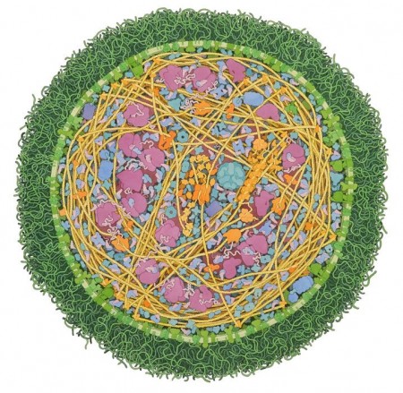
Illustration by David S. Goodsell, the Scripps Research Institute

This amazing watercolour painting shows an entire Mycoplasma mycoides cell, a bacterium that causes lung disease in cows, is painted with a brilliant green membrane that brings grass to mind. Inside, bright yellow DNA curls next to protein-builders in purple and blue. In life, this bacterium is about 250 nanometres in diameter. Click on the link to identify the membrane proteins, enzymes and other protein synthesis organelles. How beautiful is this?!
Read more about Art and Science here.

Paramecium, up close and personal!

Photo Source

This beautiful image is of a Paramecium (a common unicellular organism found in freshwater environments), using enhanced phase-contrast microscopy. It is an example of a protozoan – that is, a ‘first animal’. If you look closely, you can see the cilia (tiny, hair-like projections) that propel the organism through the water and the phospholipid bilayer (cell membrane) that is the barrier between the external and internal environment of the cell. In the top right of the image you can see water being ejected from the contractile vacuole, which helps to maintain the correct water balance inside the cell (osmoregulation). Water will naturally diffuse into the cell, as the cell’s contents have a higher concentration of salts and nutrients. The contractile vacuole is a specialized structure that collects this excess water and expels it from the cell, to prevent the cell membrane from rupturing.